Arteriography: What Is It And What Is It Used For?
Today there are multiple tests for the study of blood vessels, however, one of the most widely used is arteriography or angiography. We explain it to you.
Blood vessels form an interlocking network of pathways that are distributed through all organs of the body carrying oxygen and nutrients. The study of this system allows to guide and define the medical diagnosis many times. Arteriography is one of the most widely used methods for this purpose.
When speaking of angiography or arteriography, we refer to a paraclinical radiological study that allows evaluating the state of the arteries and any alteration in them. It uses a contrast medium instilled in the vessel that allows it to color and define the elements inside any artery.
Currently there are a wide variety of options to visualize blood vessels, among which we mention Doppler ultrasound and computerized axial tomography (CT). However, angiography is used when it is necessary to see with great detail and sharpness.
When can it be needed?
By evaluating the patient, the doctor can identify signs and symptoms that point to a condition or damage to the interior of a blood vessel. The most common reason for an angiography usually be l to clinical suspicion of blockage or occlusion of the lumen of an artery.
Manifestations such as weakness, blurred vision, headache, discomfort when walking or loss of strength in a part of the body could be the result of an arterial blockage. In these cases, angiography can identify the location and cause of said occlusion, as well as offer characteristics that facilitate the definition of severity.
Among the conditions that can be diagnosed using angiography, the following stand out:
- Aneurysms or pathological arterial dilations with risk of rupture.
- Arteriovenous malformations (AVM) or abnormal connections between vessels.
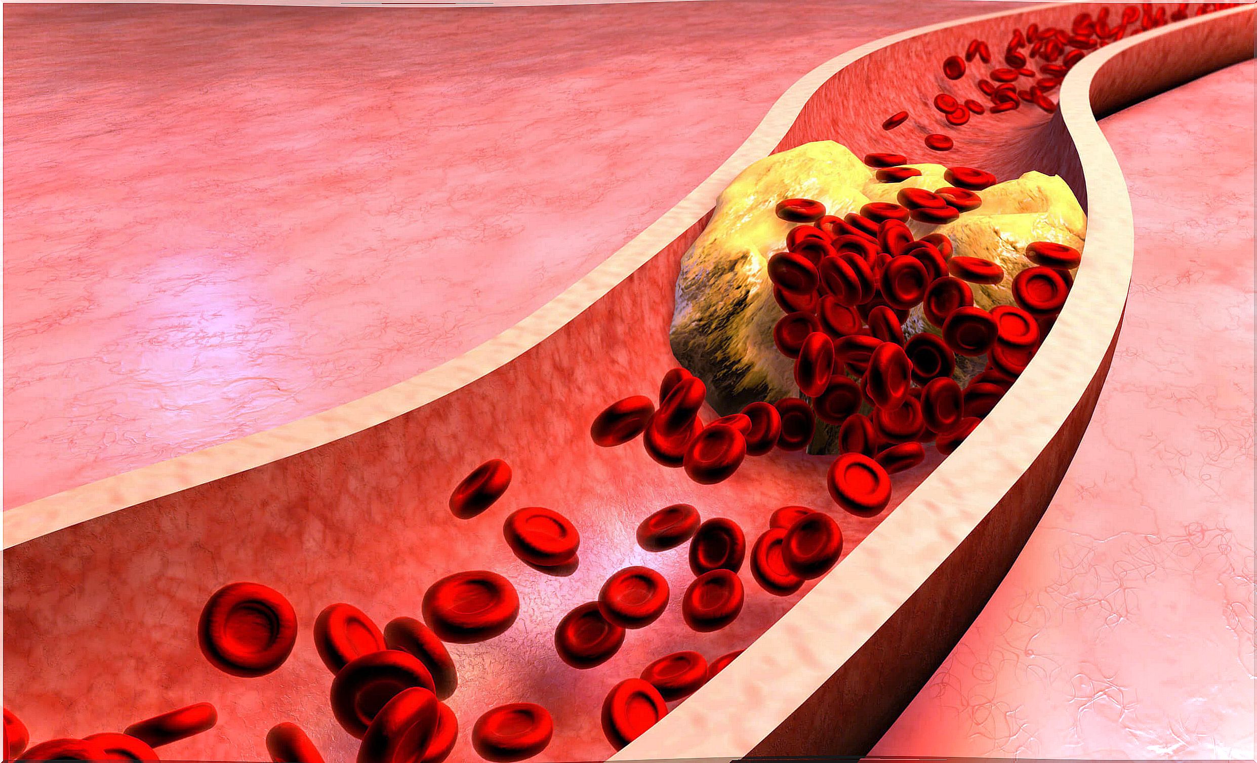
- Atherosclerotic plaques.
- Renal blood flow disorders.
- Vascular injuries after trauma.
- Dissection of the aorta.
- Clots and emboli.
Similarly, this radiological method can be used to guide the surgeon during blood vessel reconstruction procedures. It is considered the standard in the assessment of the anatomy of the coronary arteries.
What types of arteriography are there?
Angiography allows the evaluation of any blood vessel in the body, from the small arteries of the heart and brain to the large vessels of the lower limbs. So it is classified according to the organ or area to be evaluated.
Coronary arteriography or coronary angiography
It is a minimally invasive method that offers detailed images of the interior of the coronary arteries, which originate from the ascending aorta and supply the cardiac muscles. It is performed with the aim of diagnosing arteriopathies and heart disorders.
Similarly, this procedure is indicated in the following situations:
- Define the need for coronary artery bypass grafting or angioplasty.
- Evaluate the severity and degree of a myocardial ischemic pathology.
- Determine the origin of chest pain associated with respiratory distress.
- Identify the properties and characteristics of the heart prior to reconstruction of the heart valves.
Aortic arteriography or aortography
It is used with the aim of exploring and evaluating the blood flow of the aorta, from its birth in the heart to its thoracic and abdominal route.
The search includes the primary detection of pathological lumps and dilations, as well as the identification of aortic valve failures.
Similarly, it has shown great utility in the detection of aortic wall ruptures or dissection with an identification capacity of more than 50% of cases.
Cerebral arteriography
Cerebral angiography is indicated in the evaluation of alterations located within the small blood vessels of the brain. Its main utility lies in the diagnosis of narrow or blocked vessels when ischemic events that define a cerebrovascular accident are suspected.
This method is useful in identifying abnormal connections between cerebral arteries and veins. In addition, it allows to evaluate the inflammation of the wall of the blood vessels or vasculitis.
Other types of arteriography
The types of arteriography described are usually the most frequently used. However, the following are also worth noting:
- Of limbs, arms or legs.
- Fluorescein angiography for ocular evaluation.
- Pulmonary.
- Renal arteriography.
- Mesenteric angiography for exploration of the colon and small intestine.
- Pelvic
How to perform the exam?
This paraclinical tool is known as catheterization because of the way the exam is performed. It is specified with interventional or vascular radiologists.
The method consists of placing a small plastic tube or catheter in the line or blood vessel that needs to be explored. Then an iodinated contrast medium is injected to enhance the internal structures.
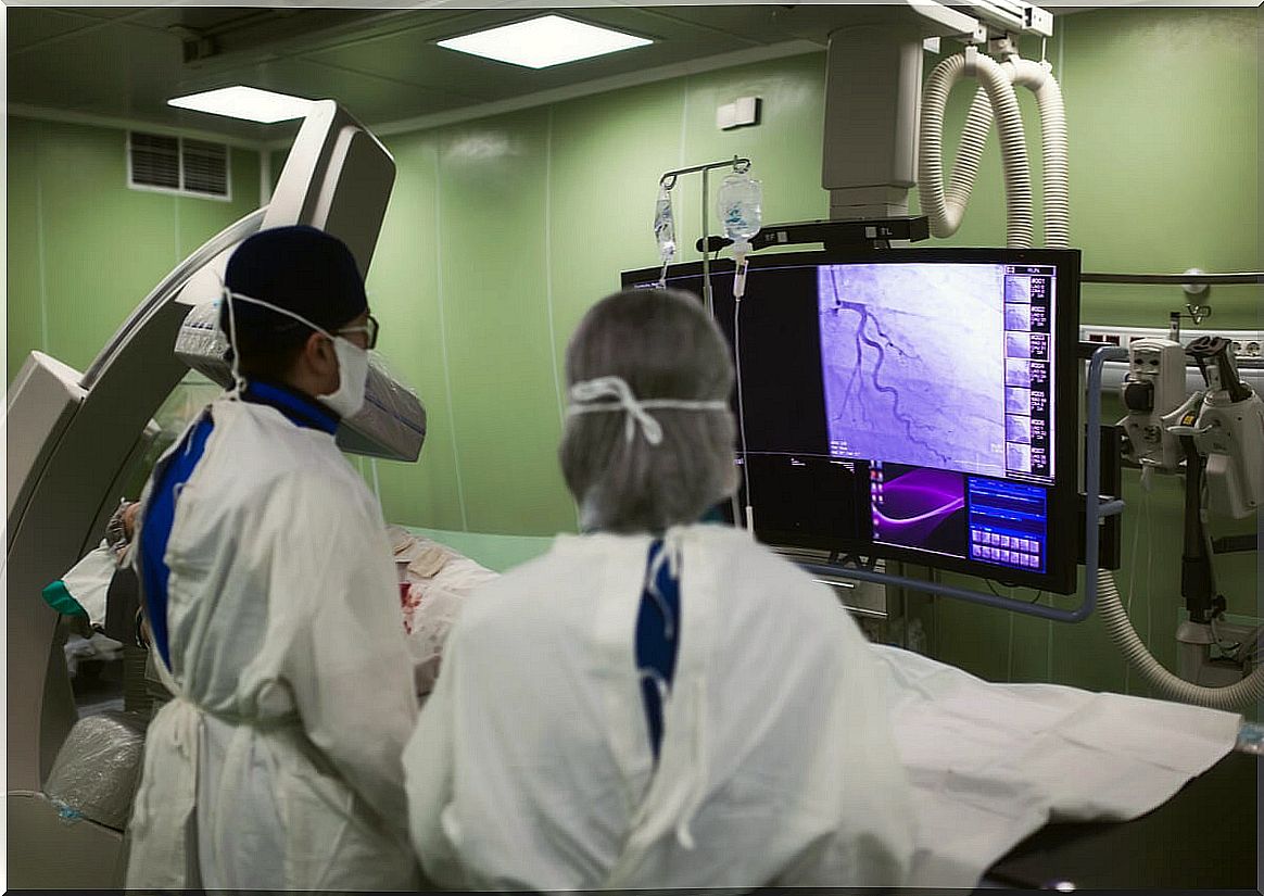
Once the contrast is circulating in the blood, several continuous radiographs are generated that allow the flow of blood through the vessel to be easily visualized in real time.
Preparation prior to an arteriography
Angiography is an invasive exploratory method that requires several preparation criteria before it, with the aim of avoiding complications in the patient and ensuring success. Some guidelines to keep in mind are the following:
- Medicines : it is vital to inform the doctor about the consumption of any medicine, especially if anticoagulant drugs or insulin are used.
- Allergic reactions : any allergy related to drug use should be reported to the specialist in the days prior to the study, especially if an allergy to X-ray contrast has been diagnosed.
- Food : it is advisable not to consume any solid or heavy food after midnight before the study.
- Smoking habit: not using cigarettes or tobacco in the previous 24 hours.
- Pregnancy : notification of pregnancy is mandatory, considering that radiation could be harmful to the baby.
- Lactation : if you are breastfeeding a newborn you should inform the specialist. Similarly, it is advisable to express and store the milk in advance. However, the American Radiological Association reported that the amount of contrast that can pass to the fetus is minimal.
How is the arteriography procedure?
Arteriography is a minimally invasive process that usually takes about 90 minutes, depending on the area to be studied and the complications that may occur. Generally speaking, the whole process can be summarized in 3 phases.
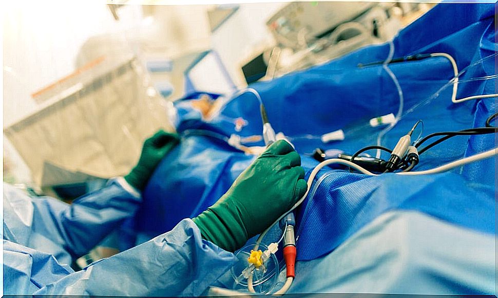
1. Introduction of the catheter
Once all the relevant preparations have been made, the specialist in charge will guide you and lay you down on an X-ray table. Once you are lying down , the area where the catheter will be inserted will be cleaned.
The specialist in charge should place local anesthesia in the area of introduction to avoid discomfort. You will then proceed with the insertion of the catheter.
In most cases it is inserted through the groin and a small incision is usually necessary. However, introduction can also be done through the arm or wrist.
2. Catheter location and contrast injection
When the catheter is in a large-caliber artery, the specialist will proceed to move it through its path until the segment to be studied is located. It is important to note that at this point you will not perceive any type of discomfort from the local anesthesia.
At the time of locating the segment to be studied, the specialist will proceed to inject the contrast medium, which will be in charge of staining the arteries.
While the contrast is being instilled, multiple plates must be taken to be able to study all the paths. This is a process that can take several minutes and successive images are necessary.
3. Catheter removal
Once all the necessary images are taken to study the path, the catheter must be removed. Removal of the same does not usually cause pain. It will only be necessary to maintain constant pressure on the area for about 20 minutes to prevent bleeding.
Results and recovery from an arteriogram
The end result of an angiography is the complete visualization of the course of the arteries in a certain area. The exam will be normal when the contrast stains all the vessels evenly.
Among the abnormal results that can be found in this study are the following:
- Contrast leakage into the extravascular medium, indicating internal bleeding.
- Narrow or blocked arteries.
- Blood vessels in abnormal positions.
Recovery is quick in most cases. The hospital stay is usually less than 6 hours in outpatients. Medical personnel should only be alert to the presence of bleeding at the catheter insertion site.
Among the general recommendations that people should follow are those to rest for 24 hours, not walk a lot and avoid smoking.
Possible complications of an arteriogram
As it is an invasive imaging study, the complications that may occur are related to both the contrast medium and the introduction of the catheter itself.
However, they are rare and the following stand out:
- Bleeding during the hours after the study.
- Allergy to contrast medium, with allergy to iodine being a common condition.
- Formation of a blood clot in the incision and subsequent embolism.
- Damage or perforation of an artery when maneuvering the catheter.
- Infection of the incision site.
An ideal study to evaluate the arteries
Arteriography is a paraclinical exam that shows a detailed image that allows an accurate analysis of the condition of the blood vessels. Although it is an invasive process, complications are rare and are not life threatening when detected early.
Recovery is quick in most cases and does not usually impede daily activities for a long time. However, it is recommended to rest and not make excessive efforts for at least 24 hours.